EYE EXAMINATIONS
Eye examinations measurements to calculate the correct intraocular lens
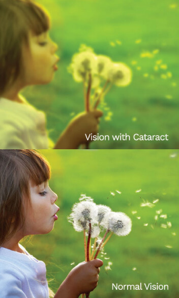
Cataract is the clouding of the natural lens in our eye that may lead to blurring of vision. No-Blade Cataract Surgery, also known as Femtosecond Laser-Assisted Cataract Surgery (FLACS) uses a Femtosecond laser (a powerful light beam) to assist in removing the cataract. This technology gives more surgical precision and safety in cataract surgery.
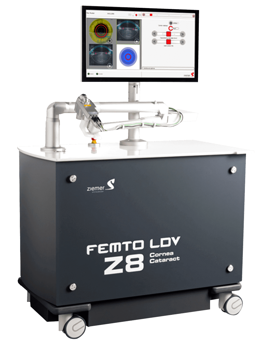
The Z8 is the first mobile femtosecond laser for refractive and cataract surgery. This innovative laser provides a more powerful laser source allowing more flexibility without losing precision.

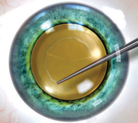
The laser places tiny, precisely shaped and positioned tubes (openings) on the eye.
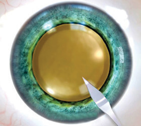
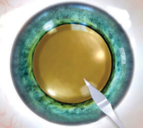
A scalpel or a diamond blade is used to make an opening on the eye.
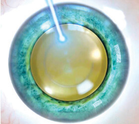
Using laser light , a perfectly sized, circular & centered opening into the lens capsule is done.
The lens capsule is manually opened, using a metal hook or forceps.
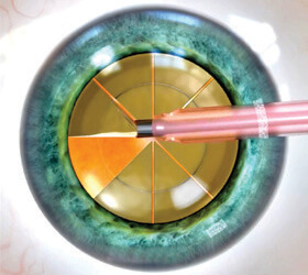
The laser fragments the clouded lens into precise sections within seconds.

The ultrasound fragments the clouded lens using an ultrasound wand.
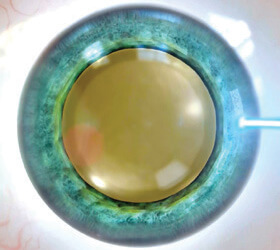
The fragmented lens is removed by aspiration and irrigation. The artificial lens is inserted with an injector.
The fragmented lens is removed by aspiration and irrigation. The artificial lens is inserted with an injector.
The most common type of intraocular lens has a monofocal optic with a single corrective power (focal point). Monofocal lenses are designed to provide clear vision for one distance, usually far, which is important for tasks such as driving. However, patients with monofocal IOLs may continue to need eyeglasses for activities at other distances, for example, reading.
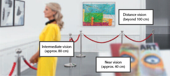
Vision simulation with a monofocal lens: clear vision at distance
Similar to bifocal eyeglasses, bifocal intraocular lenses have two focal points to provide clear vision for both distance and up close. Patients treated with bifocal IOLs may need to wear eyeglasses for certain intermediaterange tasks (approx. 80 cm) such as computer work.
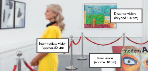
Vision simulation with a bifocal lens: clear near and distance vision
The most advanced intraocular lenses today, trifocal IOLs, have been used to successfully treat cataracts for several years. These lenses are designed to project multiple images onto the retina, which the brain combines into one sharp picture, allowing patients to see objects clearly at various distances – similar to progressive eyeglass lenses. Trifocal intraocular lenses are designed to enable not only clear far vision and a comfortable reading distance. This type of IOL provides in addition good intermediate vision, which is essential for performing daily activities such as cooking or computer work. As a result, many patients with trifocal lenses no longer need to wear glasses.
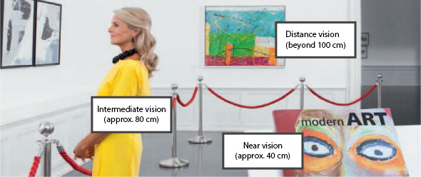
Vision simulation with a trifocal lens: clear near, intermediate and distance vision
Step 1: Corneal Incision
Starting with the corneal incision, the ophthalmologist (eye surgeon) uses a metallic blade to create the incision that allows access to the inside of the eye. When using femtosecond laser in this step we decrease the risk of inaccuracy, bleeding and inflammation allowing for a better healing process. With the help of 3D technology and the use of femtosecond laser, the surgeon can perform the incision with highest standards of accuracy. This allows the incision to self-heal and decrease the risk of inflammation and infection.
Step 2: Capsulotomy
Through the incision, the surgeon reach the clouded lens by opening the capsule that hold it in place. When using femtosecond laser in this step, we allow minimal damage to surrounding tissue! Because we need the remainder of the lens capsule to hold the artificial intraocular lens in place forever. Also the use of femtosecond laser in this step allows better stability positioning and centering of the IOL.
Step 3: Cataract Fragmentation!
Using femtosecond laser in this step (Femto-cataract) allows the surgeon to breaks up the cataract lens into smaller pieces. So less energy is used to remove it thus decreasing the risk for incision distortion!
Eye examinations measurements to calculate the correct intraocular lens
Same-day procedure with a local anesthetic such as eye drops
First day after surgery, then for approx. a month, as needed
Periodic check-ups by your eye doctor

General Ophthalmologist, Refractive Surgeon

Corneal Specialist, Refractive Surgeon

General Ophthalmologist, Refractive Surgeon

General Ophthalmologist, Refractive Surgeon

General Ophthalmologist

Retinal Specialist, Refractive Surgeon

General Ophthalmologist, Refractive Surgeon

General Ophthalmologist

General Ophthalmologist

General Ophthalmologist, Refractive Surgeon

General Ophthalmologist, Refractive Surgeon
Optimax Eye Specialist Centre-TTDI, KL (HQ)
Unit 2-2-1, Bangunan AHP,
Jalan Tun Mohd Fuad 3,
Taman Tun Dr. Ismail, 60000 K.L.
Optimax Eye Specialist Hospital-Penang
223, Jalan Masjid Negeri,
11600 Penang.
Optimax Eye Specialist Centre-Bandar Sunway
No. 11 Ground Floor,
Jln PJS 11/28B, Sunway,
46150 P.J., Selangor
Optimax Eye Specialist Centre-Sri Petaling
No.145 Jalan Radin Bagus,
Sri Petaling,
57000 Kuala Lumpur.
Optimax Eye Specialist Centre-Klang
No. 17, Jalan Bayu Tinggi 7,
41200 Klang,
Selangor.
Optimax Eye Specialist Centre-Shah Alam
50G, Blok 3, Jalan Pahat G15/G,
Dataran Otomobil, Seksyen 15,
40200 Shah Alam.
Optimax Eye Specialist Centre-Seremban
No. 141, Jalan Tun Dr. Ismail,
70200 Seremban,
Negeri Sembilan.
Optimax Eye Specialist Centre-Johor Bahru
53 & 55, Jalan Cantik 6,
Taman Pelangi Indah,
81800 Ulu Tiram, Johor.
Optimax Eye Specialist Centre-Muar, Johor
1-5 & 1-6,
Jalan Ismail, Muar,
84000 Johor.
Optimax Eye Specialist Centre-Kluang
No.43 & 44, Jalan Haji Manan,
86000 Kluang,
Johor.
Optimax Eye Specialist Centre-Segamat
49B & 49C, Jalan Genuang,
85000 Segamat,
Johor.
Optimax Eye Specialist Centre-Ipoh
No.1 Jalan Dato Khong Kam Tak,
Off Jalan Tambun, 31400 Ipoh,
Perak
Optimax Eye Specialist Centre-Kuching
No 59 & 61, Ground Floor,
Jalan Tun Jugah,
93350 Kuching, Sarawak.
Services offered : Eye Examination, Cataract surgery, Laser Vision Correction (LASIK), Implantable Collamer Lens (ICL), Refractive Lens Exchange (RLE), Management & Treatment of Eye Diseases Paediatric (Children) Eye Care, Corneal Reshaping Therapy (Ortho-K)